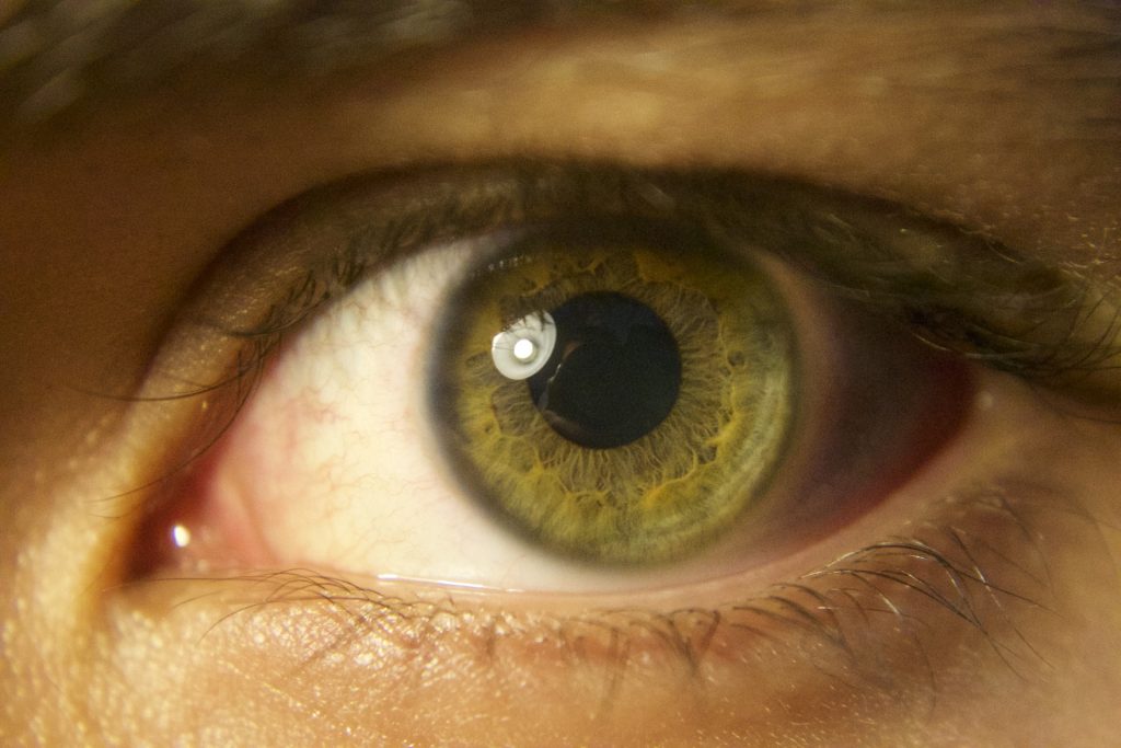
Dr. Peter Snyder, a professor of Biomedical Sciences at the University of Rhode Island, has been conducting research into retinal scanning technology that could detect signs of Alzheimer’s as early as 20 years before symptoms appear. Scientists are interested in the retina because it is an extension of the brain. As part of our nervous system it contains the same neurons, glial cells, vascular supply, and chemistry as our brains do.
The retina can provide clues about possible Alzheimer’s, and because it is exposed tissue it can be examined in microscopic detail without causing harm or discomfort, and without being invasive. This procedure is significantly less expensive than brain scans, and older adults often see ophthalmologists regularly.
Through retinal scans these researchers found a layer of cells showing early changes, in the pre-clinical stages of the disease before the onset of symptoms, providing evidence of increased volume in the structure, which they think represents an inflammatory process. Inflammation in the brain is a key component in the development of Alzheimer’s disease, and there is some evidence of that in the retina. Their study revealed something they are calling occlusion bodies which they suspect are blotches of tissue that contain beta-amyloid protein, and that are interfering with the transmission of light from the scanner.
They are trying to develop a screening test to know when older adults need to be referred to a specialty center for more diagnostic tests. A screening test is not the same as making a confirmatory diagnosis; there is generally some false/positive error in these tests to catch more people who may have the disease, rather than taking the chance of missing those who may go on to have the disease.
An ophthalmologist may notice changes in a patient’s retina that would indicate a need to refer him or her to a neurologist or a memory disorder center in order to get more testing. The goal is to intervene earlier than has been done in the past. Diagnosing Alzheimer’s earlier is better, becauseonce someone is clinically showing symptoms, the disease has already been active for one or more decades and may have caused irreversible damage to the brain. The theory is that developing effective therapies to slow the progression of Alzheimer’s may not be possible until there is a means of diagnosing and intervening earlier than is currently possible.
Dr. Snyder reported that the last study they finished (the five-year trial) gave them some evidence that they are on the right path. Their next trial is to confirm it, and to determine a method for scoring the retinal imaging that can be translated into a test which can be used by optometrist and ophthalmologists.
Hi Linda,
This is Jim Lukanich ‘75, an old acquaintance from Ripon days! I was catching up on Jondi’s class newsletters and saw your update (wow – actually from a year ago!). I obviously connected to your blog post as a result and was particularly interested in this piece.
A couple of years ago I was applying for additional life insurance and ran into an interesting issue with the insurance company’s approval. Although I qualified for their top health rating, they did make me jump through a number of “hoops” before approving my policy. It turns out that because the only medication I was taking, and am still taking, is latanprost eye drops for glaucoma, they needed to dig more deeply into my family’s history with dementia and alzheimer’s disease. It was at this time that I found out about the medical community’s connection of glaucoma with the development of the optic nerve during gestation and alzheimer’s disease.
Congratulations on your work on this area and needless to say I’ll be buying your book, assuming I remember!